 By Atlantic Endo Medical Review Board
More than half of all adults will develop a thyroid nodule at some point in their lives, yet the vast majority will never need surgery. Comprehending thyroid nodules: when to watch and when to worry is one of the most important conversations you can have with your endocrinologist, because the answer is rarely as alarming as a new diagnosis might feel.
A nodule is simply an abnormal growth of cells within the gland, that small butterfly-shaped gland at the base of your neck. Most nodules are discovered incidentally during an imaging scan ordered for an entirely different reason. While the word “nodule” can sound frightening, current medical guidelines are clear: the overwhelming majority are benign, and modern risk-based evaluation tools now allow physicians to distinguish, with remarkable precision, which nodules deserve close watching and which require more urgent action.
Table of Contents
Key Takeaways
- Most nodules are benign and do not require surgery or aggressive treatment.
- Ultrasound-based risk stratification systems guide decisions about biopsy and surveillance.
- Growth rate, nodule characteristics, and symptom burden, not size alone, determine the level of concern.
- Active surveillance is now an accepted and often preferred strategy for low-risk and even some small suspicious nodules.
- Partnering with an experienced endocrinologist ensures your nodule is evaluated with the most current, evidence-based approach.
What Is a Thyroid Nodule and Why Are They So Common
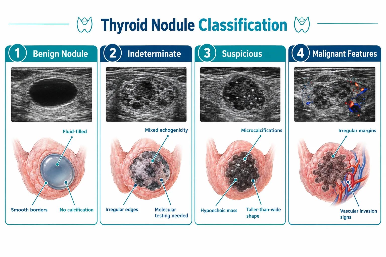
Thyroid nodules form when tissue grows in an abnormal cluster. They can be solid, fluid-filled (cystic), or a mixture of both. Some produce excess thyroid hormone; most do not. They range from microscopic to several centimeters in diameter.
Their prevalence is striking. High-resolution ultrasound detects nodules in 19 to 68 percent of randomly selected adults, with higher rates in women and older individuals. Despite this frequency, thyroid cancer accounts for only a small fraction of all nodules, roughly 5 to 15 percent, depending on the population studied and the risk factors present.
Common reasons nodules form include:
- Iodine deficiency or fluctuation
- Hashimoto’s thyroiditis (autoimmune disease)
- Multinodular goiter
- Thyroid cysts
- Benign adenomas
- Rarely, thyroid cancer
Knowing what a thyroid nodule looks like on ultrasound is the first step toward accurate risk classification, and accurate classification is what separates unnecessary worry from appropriate action.
Thyroid Nodules: When to Watch and When to Worry, The Core Framework
The central question every patient asks is simple: does my nodule need treatment, or can I safely monitor it? The answer depends on a structured evaluation that combines ultrasound imaging, laboratory tests, and sometimes a fine-needle aspiration (FNA) biopsy.
Ultrasound Risk Stratification
Modern endocrinology relies on validated ultrasound scoring systems, most notably the American College of Radiology’s TI-RADS (Thyroid Imaging Reporting and Data System) and the American Thyroid Association (ATA) guidelines, to assign risk categories to every nodule. These systems evaluate:
| Ultrasound Feature | Lower Risk | Higher Risk |
|---|---|---|
| Composition | Purely cystic or spongiform | Solid or predominantly solid |
| Echogenicity | Isoechoic or hyperechoic | Hypoechoic or markedly hypoechoic |
| Shape | Wider than tall | Taller than wide |
| Margins | Smooth, well-defined | Irregular, lobulated, or extrathyroidal |
| Calcifications | None or peripheral | Microcalcifications |
A nodule that scores low on these features is assigned to a “watch” category. One that scores high triggers a recommendation for biopsy. This is not a guessing game, it is a data-driven process. You can learn more about how suspicious-looking nodules on thyroid sonogram are assessed for malignancy to better cmprehend how radiologists and endocrinologists interpret these findings together.
When “Watch and Wait” Is the Right Answer
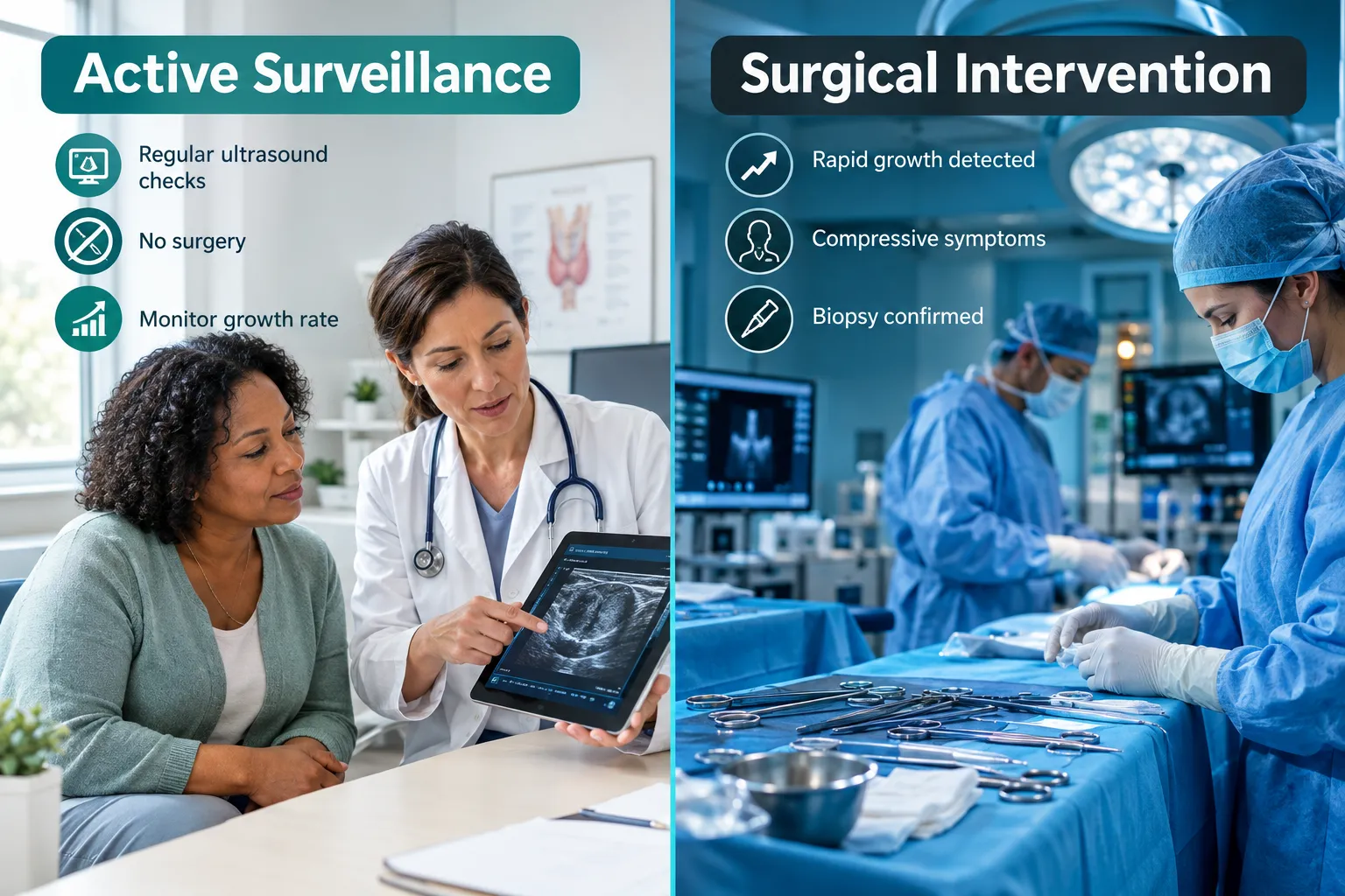
For nodules classified as very low risk or low risk, clinical guidelines endorse active surveillance, meaning scheduled ultrasound follow-up without biopsy or surgery. This approach is appropriate when:
- The nodule is purely cystic or spongiform
- The nodule is smaller than 1 cm with no suspicious ultrasound features
- The nodule has been stable in size over prior imaging studies
- The patient has no high-risk clinical history (prior radiation exposure, family history of cancer)
Active surveillance is not passive neglect. It is a deliberate, structured monitoring plan. Repeat ultrasounds are typically performed at 6 to 12 months after the initial finding, then at 12 to 24-month intervals if stability is confirmed.
Red Flags That Shift the Balance Toward Action
Certain findings move a nodule from the “watch” column to the “worry” column. These include:
- Rapid growth: A 20 percent or greater increase in at least two nodule dimensions, or more than 2 mm growth in two dimensions, within a surveillance period
- New or worsening symptoms: Difficulty swallowing, a sensation of pressure in the throat, hoarseness, or a persistent cough
- Suspicious biopsy results: Bethesda categories IV, V, or VI on cytology
- Lymph node changes: Enlarged or structurally abnormal cervical lymph nodes on ultrasound
If you are experiencing any of these symptoms alongside a known or suspected nodule, this is the moment to seek evaluation promptly rather than waiting for a scheduled appointment. Our detailed resource on thyroid nodules, when to worry, what to watch, and when to act provides an expanded look at these clinical thresholds.
The Role of Biopsy, Molecular Testing, and Active Surveillance
When ultrasound findings are suspicious enough to warrant a biopsy, fine-needle aspiration (FNA) is the standard procedure. It is minimally invasive, typically performed in an outpatient setting with ultrasound guidance, and provides cellular material for laboratory analysis.
Biopsy results are reported using the Bethesda System, a six-category classification that ranges from non-diagnostic (Category I) to malignant (Category VI). Categories III and IV, often called “indeterminate”, represent a significant clinical challenge because they are neither clearly benign nor clearly cancerous.
Molecular Testing: A Game-Changer for Indeterminate Nodules
For indeterminate nodules, molecular testing panels (such as Afirma Gene Sequencing Classifier or ThyroSeq) analyze the genetic material within biopsy cells to refine the risk estimate. This technology has meaningfully reduced the number of patients who undergo diagnostic surgery only to find a benign nodule.
Importantly, current guidelines are shifting toward active surveillance rather than automatic surgery for certain indeterminate nodules with low-risk molecular profiles, particularly those with RAS-type mutations, which tend to behave indolently. This represents a significant evolution in thyroid nodule management and reflects a broader principle: that quality of life matters as much as cancer detection.
Expanding the Role of Active Surveillance
Active surveillance was originally reserved for papillary microcarcinomas (tumors smaller than 1 cm). Evidence now supports extending this approach to selected patients with small, highly suspicious nodules, provided they are followed by an experienced specialist in a structured program.
Main criteria for active surveillance candidacy typically include:
- Tumor size under 1 to 1.5 cm
- No evidence of lymph node involvement or distant spread
- No tumor abutment of the trachea or recurrent laryngeal nerve
- Patient preference and ability to adhere to follow-up
Studies consistently show that quality of life outcomes are favorable with active surveillance compared to immediate surgery for appropriately selected patients. The risk of disease progression during surveillance is low, and surgical rescue, when needed, remains effective.
To know the full diagnostic picture, including what your imaging results mean, explore our guide on ultrasound for thyroid in New York and learn what to expect at your first endocrinologist thyroid visit.
Nodules: When to Watch and When to Worry in Special Populations
Certain groups require modified surveillance strategies or heightened vigilance:
Children and adolescents: Thyroid nodules in pediatric patients carry a higher malignancy rate than in adults. Biopsy thresholds are lower, and surgical consultation is more frequently recommended.
Pregnant women: Nodules discovered during pregnancy are evaluated with ultrasound. FNA can be safely performed during pregnancy if findings are concerning. Radioactive iodine scans are contraindicated. A thyroid ultrasound is safe during pregnancy and remains the primary imaging tool.
Patients with prior radiation exposure: A history of head and neck radiation significantly increases malignancy risk. Lower biopsy thresholds and more frequent surveillance intervals apply.
Patients with a family history of this cancer or MEN syndromes: Genetic counseling and earlier intervention may be warranted.
How Atlantic Endocrinology Approaches Thyroid Nodule Care
At Atlantic Endocrinology & Diabetes Center, thyroid nodule evaluation is a core specialty. Dr. Stella Ilyayeva, M.D., our lead endocrinologist with over 20 years of clinical experience, leads a team that integrates on-site thyroid ultrasound, laboratory services, and multidisciplinary consultation, including interventional radiology with Dr. Iftikhar Ahmad, who brings over 25 years of experience in minimally invasive procedures.
Our approach is built on the principle that every patient deserves a personalized, risk-based evaluation, not a one-size-fits-all protocol. We use current ATA and TI-RADS guidelines, molecular testing when appropriate, and shared decision-making to help you know your options clearly.
We serve patients across Queens and Brooklyn from multiple convenient locations:
- Forest Hills: 99-45 67th Rd Suite 103, Forest Hills, NY 11375
- Rego Park: 97-25 63 Dr 1 FL, Rego Park, NY 11374
- Bushwick/Brooklyn: 359 Stockholm Street, Brooklyn, NY 11237
We accept a broad range of insurance plans including Medicare, Medicaid, Aetna, United Healthcare, EmblemHealth, Healthfirst, Cigna, and many others.
Frequently Asked Questions
Can a nodule go away on its own?
Some thyroid nodules, particularly purely cystic ones, can shrink or resolve without treatment, especially if they are related to temporary inflammation or fluid accumulation. Nevertheless, solid nodules rarely disappear on their own. Rather than waiting to see if a nodule resolves, it is important to have any newly discovered nodule evaluated by an endocrinologist so that an appropriate monitoring or treatment plan can be established.
How often should a nodule be monitored with ultrasound?
Monitoring frequency depends on the nodule’s risk category. Low-risk nodules are typically re-imaged at 12 to 24 months after the initial evaluation, then less frequently if stability is confirmed. Intermediate or high-risk nodules, or those that have shown growth, may require follow-up at 6 to 12-month intervals. Your endocrinologist will determine the schedule based on your individual findings and risk profile.
Does having a thyroid nodule mean I have thyroid cancer?
No. The vast majority of thyroid nodules are benign. Depending on the population studied, only approximately 5 to 15 percent of nodules evaluated by biopsy are found to be malignant. Even among those, many are slow-growing, highly treatable forms of cancer with excellent long-term outcomes. A thorough evaluation, including ultrasound and, when indicated, biopsy, is the only reliable way to determine the nature of a specific nodule.
What symptoms should prompt me to call my doctor about a thyroid nodule right away?
Contact your physician promptly if you notice rapid enlargement of a neck lump, difficulty swallowing or breathing, persistent hoarseness or voice changes, pain in the front of the neck, or swollen lymph nodes. These symptoms do not automatically indicate cancer, but they do warrant urgent evaluation to rule out serious causes and guide appropriate management.
References
- Haugen, B. R., et al. (2016, updated with 2023 revisions). American Thyroid Association Management Guidelines for Adult Patients with Thyroid Nodules and Differentiated Thyroid Cancer. American Thyroid Association. https://www.thyroid.org/professionals/ata-professional-guidelines/
- Tessler, F. N., et al. (2017, reaffirmed 2022). ACR Thyroid Imaging, Reporting and Data System (TI-RADS): White Paper of the ACR TI-RADS Committee. Journal of the American College of Radiology. https://www.acr.org/Clinical-Resources/Reporting-and-Data-Systems/TI-RADS
- National Cancer Institute. (2023). Thyroid Cancer Treatment (PDQ), Patient Version. U.S. National Institutes of Health. https://www.cancer.gov/types/thyroid/patient/thyroid-treatment-pdq
- Ito, Y., et al. (2021). Active surveillance as the first-line management for low-risk papillary thyroid microcarcinoma. European Journal of Surgical Oncology, 47(2), 283-291. https://doi.org/10.1016/j.ejso.2020.09.008
- American Thyroid Association. (2022). Thyroid Nodule Patient Information. https://www.thyroid.org/thyroid-nodules/
- Brito, J. P., et al. (2021). Thyroid cancer: zealous imaging has increased detection and treatment of low risk tumours. BMJ, 373, n1240. https://doi.org/10.1136/bmj.n1240
- National Institute of Diabetes and Digestive and Kidney Diseases. (2021). Thyroid Nodules. U.S. Department of Health and Human Services. https://www.niddk.nih.gov/health-information/endocrine-diseases/thyroid-nodules
Medical and Editorial Commitment
The content of this article is strictly for educational and informational purposes. It does not replace in-person professional medical diagnosis, advice, or treatment..
Read Privacy PolicyRelated Posts
What Do the Adrenal Glands Actually Do? A Patient’s Guide
Two walnut-sized glands sitting atop your kidneys are quietly running some of the most critical operations in your body, and most people [...]
The Complete Diabetes Check Guide: Tests, Timing, and What Your Results Mean
More than 38 million Americans are living with diabetes today, and roughly one in five of them do not know it yet. [...]