
 By Atlantic Endo Medical Review Board
Seeing a suspicious-looking nodule on your thyroid sonogram can immediately raise the question: Is it cancer? Many patients jump to the worst conclusion, assuming that any feature hinting at malignancy is a guaranteed diagnosis. The visual evidence on an ultrasound is rarely a final verdict. If you are seeking reliable Ultrasound services in Queens, NY, you must ask: Are All Suspicious Looking Nodules on Thyroid Sonogram Malignant? The surprising answer is inside.
TL;DR
Despite suspicious features, 95% of thyroid nodules are benign. TI-RADS uses ultrasound criteria—composition, margins, and microcalcifications—to assess risk. Biopsy (FNA) is only recommended when multiple high-risk signs accumulate, as ultrasound is a risk map, not a final diagnosis.
Continue care through Ultrasound services in Queens, NY
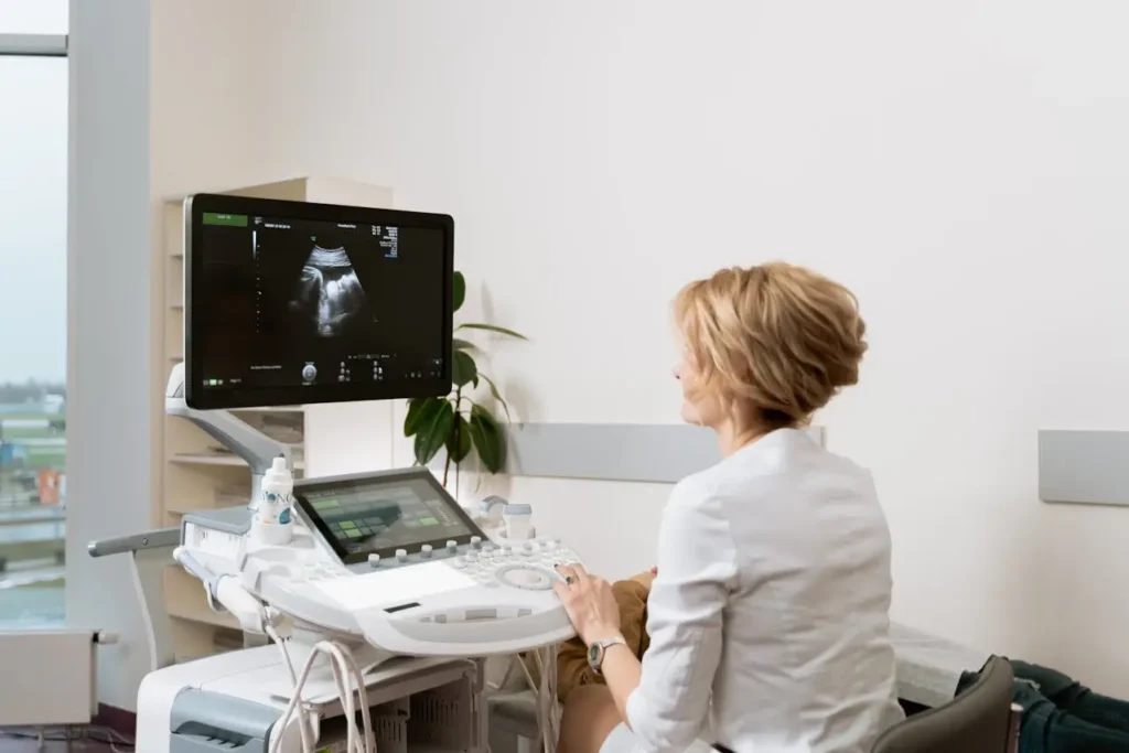
Table of Contents
What does a Thyroid Nodule Look Like on Ultrasound?
On an ultrasound, a thyroid nodule appears as a “spot” inside the gland, differentiated from healthy tissue by its composition, brightness, and edges. It can be cystic (black and fluid-filled), solid, or mixed, with variations in its appearance based on these characteristics.
Echogenicity aids in its assessment: hypoechoic nodules are darker and can be more suspicious, while hyperechoic ones are usually brighter and normally benign. Margins are also analyzed, with irregular or spiculated margins being more concerning than defined ones.
Other factors include shape (taller-than-wide), the presence of microcalcifications, and internal vascularity. These characteristics combine to classify the risk and decide if additional studies, such as a biopsy, are required.
Ultrasound Appearance of Solid and Cystic Nodules
Thyroid ultrasound is essential for the initial assessment of any mass, as it allows for rapid distinction of whether a nodule is solid, cystic, or mixed—a differentiation necessary to determine the malignancy risk level. This compositional distinction is the first step in deciding if additional studies, such as a biopsy, are required.
- Cystic Nodules: These are observed as anechoic structures (completely black) on the ultrasound image due to their liquid content. They are normally considered benign.
- Solid Nodules: These are composed mainly of tissue and manifest as masses that can be hypoechoic (darker than the surrounding thyroid tissue) or hyperechoic (brighter). Echogenicity (brightness) is decisive for their classification.
- Mixed Nodules: These combine solid and cystic components. In these cases, assessment focuses on the characteristics of the solid component, as this is what determines the actual level of suspicion for malignancy.
Ultimately, the ultrasonic appearance of a nodule, especially one with a solid component, guides the radiologist to classify the risk and make an informed decision about the need for surveillance or fine-needle aspiration (biopsy), ensuring a precise assessment.
Nodule Shape Margins and Border Characteristics
The ultrasound assessment of thyroid nodules delves into their morphological structure. Classification systems like ACR TI-RADS use the nodule’s shape, margins, and border characteristics as indicators to determine its risk of malignancy. Observing whether the nodule is “taller-than-wide” or has irregular borders are factors that guide the need for additional studies.
| Characteristic | Finding Suggestive of Benignity | Finding Suggestive of Malignancy |
|---|---|---|
| Shape | Oval or rounded | Taller-than-wide (Non-parallel), Irregular |
| Margins | Smooth (well-defined) | Irregular, microlobulated, spiculated, Poorly defined |
| Border (Halo) | Thin and complete peripheral halo | Thick, incomplete, or irregular halo |
| Associated Findings | Comet-tail artifact (Colloid) | Extrathyroid extension, Microcalcifications |
These morphological characteristics are essential for risk classification. A nodule with an oval shape, smooth margins, and a complete halo tends to be classified as benign. Conversely, the presence of a ‘taller-than-wide’ shape, spiculated margins, or capsule invasion (extrathyroid extension) significantly increases suspicion, justifying the recommendation for a biopsy or closer monitoring.
Echogenicity Patterns Compared to Normal Thyroid Tissue
Echogenicity describes the intensity of the echo reflected by the nodule compared to normal thyroid parenchyma. This is an essential characteristic to determine a nodule’s risk. On the ultrasound, the normal thyroid gland appears homogeneous and of medium echogenicity. Nodules are classified as follows:
- Hypoechoic (Darker): Show lower echogenicity than the surrounding tissue. Marked hypoechogenicity correlates with a significantly higher risk of malignancy (primarily papillary carcinoma).
- Isoechoic (Same brightness): Have an echogenicity similar to that of normal thyroid tissue. These nodules usually carry a low or intermediate risk.
- Hyperechoic (Brighter): Present greater echogenicity. They are normally considered a low-risk finding.
The presence of marked hypoechogenicity is a significant risk factor for malignancy. However, this characteristic is always evaluated in conjunction with the shape, margins, and presence of calcifications to achieve a complete and accurate risk classification, thus guiding the decision on the need for a biopsy or follow-up.
Microcalcifications and Internal Vascularity Findings
The joint evaluation of microcalcifications and the identification of internal vascularity are ultrasound findings that significantly increase the suspicion of malignancy in a thyroid nodule. These indicators are fundamental for precise risk classification using the TI-RADS system.
- Microcalcifications (Bright Spots): These are observed as small, bright echogenic foci within the nodule without generating acoustic shadowing. Histologically, they often correspond to “psammoma bodies,” a characteristic associated with greater suspicion of malignancy.
- Internal Vascularity (Doppler Flow): This is identified via Doppler as blood flow within the nodule, especially when it is predominant in the center. Central or chaotic vascularity may suggest higher risk, as it indicates an active supply for its growth.
The identification of these characteristics via ultrasound is a determining step in risk assessment. The combination of these two high-suspicion findings prompts the specialist to assign a higher risk category, which, in turn, usually guides the recommendation for fine-needle aspiration for a definitive diagnosis.
Benign Versus Malignant Risk Feature Assessment
Thyroid ultrasound is essential for differentiating low-risk from suspicious nodules, since 95% are benign. The standardized TI-RADS system classifies these findings to determine which nodules require Fine-Needle Aspiration (FNA) and to guide clinical management.
The system evaluates multiple criteria: benign nodules are cystic, have smooth margins, and are “wider-than-tall” in shape. High-risk findings, such as solid composition, “taller-than-wide” shape, irregular margins, or microcalcifications, determine a TR1 to TR5 classification.
No single ultrasound sign is definitive for diagnosing cancer; the decision for biopsy depends on the accumulation of suspicious characteristics. FNA is recommended for solid and hypoechoic nodules larger than 10 mm, classified in high grades (TR4 or TR5) on the TI-RADS. Ultrasound functions as a risk map, not a final verdict.
Key Takeaways
- Despite a suspicious ultrasound appearance, nearly 95% of thyroid nodules are benign. Ultrasound serves as an essential, standardized “risk map” to guide clinical management, but it cannot provide a definitive cancer diagnosis on its own.
- The ACR TI-RADS system standardizes assessment by assigning scores (TR1 to TR5) based on accumulated risk features. High scores (TR4 or TR5) and a size over 10 mm typically guide the recommendation for Fine Needle Aspiration (FNA).
- Highly suspicious findings include solid composition, marked hypoecogenicity, irregular margins, and a “taller-than-wide” shape. The presence of microcalcifications and chaotic internal vascularity significantly raises the malignancy alert.
- Benign nodules are typically cystic or spongiform, with smooth margins and a “wider-than-tall” shape. Isoechoic or hyperechoic appearance and a complete halo are classic signs that suggest a low risk of malignancy.
- Ultrasound is critical for immediately differentiating the nodule’s composition (solid, cystic, or mixed). For risk classification, the radiologist focuses on the solid component and its echogenicity (brightness) as the initial determining factors.
FAQs
What is an ultrasound test for?
An ultrasound is a safe, non-invasive imaging test that uses sound waves to visualize organs, tissues, and blood flow in real time. It’s commonly used to evaluate areas like the thyroid, detect nodules or cysts, and guide further diagnostic decisions.
How long does an ultrasound take?
Most ultrasound exams take between 20 and 45 minutes, depending on the area being studied and the level of detail required. Simpler scans may be shorter, while more complex evaluations can take longer.
What diseases can be detected by ultrasound?
Ultrasound can help detect conditions such as cysts, tumors, nodules, and abnormalities in organs. It’s also useful for identifying issues like gallstones, blood flow problems, and other structural changes in soft tissues.
What are the three main types of ultrasound?
The three main types are diagnostic ultrasound (to view organs and tissues), Doppler ultrasound (to assess blood flow), and procedural ultrasound (used to guide procedures like biopsies).
Sources
- Chung, R., & Kim, D. (2019). Imaging of thyroid nodules. Applied Radiology, 48(1), 16-26.
https://cdn.agilitycms.com/applied-radiology/PDFs/Issues/AR_01-19_Chung.pdf
- Bojunga, J. (2018). Ultrasound of thyroid nodules. Ultraschall in der Medizin-European Journal of Ultrasound, 39(05), 488-511.
https://www.thieme-connect.com/products/ejournals/abstract/10.1055/a-0659-2350
You May Also Like
- How Long Does a Thyroid Ultrasound Take?
- How Much Does a Thyroid Ultrasound Cost?
- Is a Thyroid Ultrasound Safe During Pregnancy? What Expecting Mothers Should Know
Medical and Editorial Commitment
The content of this article is strictly for educational and informational purposes. It does not replace in-person professional medical diagnosis, advice, or treatment..
Read Privacy PolicyRelated Posts
How Long Are You Radioactive After a Thyroid Uptake Scan?
You’ve just taken a radioactive pill to evaluate your gland's health. But as you leave the clinic, a critical question arises: are [...]
Does Fundoscopy Tell Us About Lymphoma?
What if a simple eye exam could reveal clues about conditions far beyond your vision? Fundoscopy is often used to examine the [...]