
 By Atlantic Endo Medical Review Board
More than 90 million Americans are at high risk for serious vision loss, yet most have never had a proper retinal exam. A single fundoscopy appointment, often lasting less than 20 minutes, can detect not only eye disease but also early signs of diabetes, hypertension, and even cardiovascular conditions before symptoms appear. For patients managing chronic conditions, this exam is one of the most underutilized tools in preventive medicine.
Table of Contents
Key Takeaways
- Fundoscopy is a non-invasive eye examination that allows clinicians to view the retina, optic disc, blood vessels, and macula directly.
- It is a critical screening tool for diabetic retinopathy, glaucoma, age-related macular degeneration (AMD), and optic nerve disease.
- Pupil dilation with pharmacologic drops is typically recommended for the most thorough examination.
- Artificial intelligence is rapidly transforming fundoscopy into a powerful platform for detecting systemic diseases, including cardiovascular risk.
- Patients with diabetes, hypertension, or a family history of eye disease should schedule fundoscopy at least once per year.
What Is Fundoscopy and Why Does It Matter?
Fundoscopy, also called ophthalmoscopy or a fundus examination, is a clinical procedure in which a physician uses a lighted instrument called an ophthalmoscope to examine the interior structures of the eye. The word “fundus” refers to the back portion of the eye’s interior, which includes the retina, optic disc, macula, fovea, and the network of retinal blood vessels.
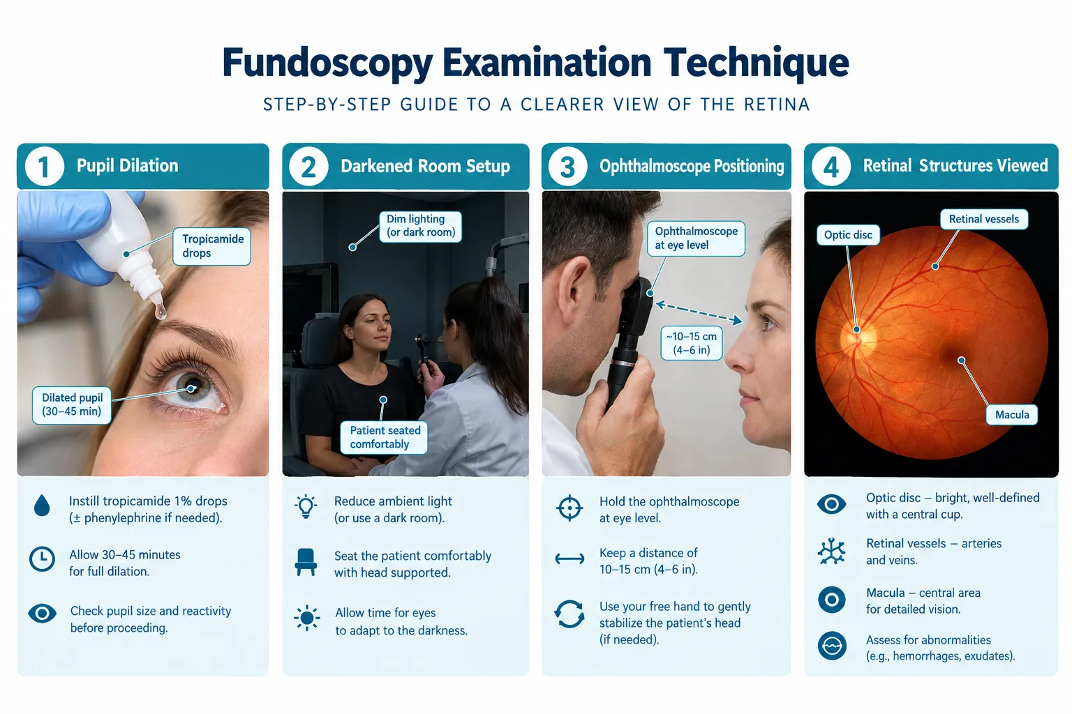
What makes this exam remarkable is its directness. The retina is the only place in the human body where blood vessels and nerve tissue can be observed without surgery or invasive procedures. Because the retinal vasculature mirrors the condition of blood vessels throughout the body, a trained clinician can gather significant systemic health information from a single look.
Structures visible during fundoscopy include:
| Structure | What It Reveals |
|---|---|
| Optic disc | Optic nerve health, signs of glaucoma |
| Retinal blood vessels | Hypertensive or diabetic vascular changes |
| Macula | Age-related macular degeneration, central vision loss |
| Fovea | Sharpness of central vision |
| Retinal periphery | Tears, detachments, tumors |
For patients working with an endocrinologist specializing in diabetes in New York, fundoscopy is often a recommended component of comprehensive annual care, given how directly uncontrolled blood sugar affects retinal health.
Types of Fundoscopy and How the Examination Works
There are three primary methods of fundoscopy used in clinical practice today.
Direct Ophthalmoscopy uses a handheld device that provides a monocular, upright image of the fundus at roughly 15x magnification. It is portable, widely available, and commonly used in primary care and emergency settings. Nevertheless, it offers a limited field of view.
Indirect Ophthalmoscopy uses a headset worn by the examiner combined with a handheld condensing lens. This method provides a wider, stereoscopic view of the peripheral retina and is preferred in ophthalmology for detecting retinal tears or detachments.
Slit-Lamp Biomicroscopy combines a slit-lamp microscope with a contact or non-contact lens to achieve a highly magnified, three-dimensional view of the posterior segment. This method is particularly useful for detailed assessment of the macula and optic disc.
Pupil Dilation: Why It Is Recommended
Most thorough fundoscopic examinations require pharmacologic pupil dilation. Eye drops, typically tropicamide, phenylephrine, or a combination, are instilled 20 to 30 minutes before the exam. Dilation widens the pupil from approximately 2-3 mm to 6-8 mm, allowing a significantly broader and clearer view of the retinal periphery.
Patients should be aware that dilation causes temporary light sensitivity and blurred near vision lasting 4 to 6 hours. Arranging transportation after the appointment is advisable.
“A dilated fundus examination remains the gold standard for detecting early diabetic retinopathy and other sight-threatening conditions before the patient notices any change in vision.”
For patients who want to understand how modern digital imaging compares to traditional methods, the resource on digital vs. traditional ophthalmoscopy and retinal imaging insights provides a detailed breakdown.
What Conditions Can Fundoscopy Detect?
Fundoscopy is a frontline diagnostic tool for a wide range of conditions. Its value extends well beyond ophthalmology into endocrinology, cardiology, and neurology.
Diabetic Retinopathy
Diabetic retinopathy is the leading cause of blindness in working-age adults worldwide. During fundoscopy, clinicians look for microaneurysms (tiny bulges in retinal vessels), hemorrhages, hard exudates, cotton-wool spots, and in advanced cases, neovascularization, the growth of fragile new blood vessels that can bleed and scar.
Early detection is critical because treatment is far more effective in the nonproliferative stages. Patients in the New York area can learn more about specialized diabetic retinopathy fundoscopy services in Queens, NY and across New York.
Regular exercise and blood sugar management directly reduce retinopathy risk, a topic explored in depth in this guide on whether exercise helps diabetes and key insights for managing the condition.
Glaucoma
Glaucoma causes progressive optic nerve damage, often without early symptoms. Fundoscopy reveals characteristic changes to the optic disc, including increased cup-to-disc ratio and notching of the neuroretinal rim, which signal nerve fiber loss.
Age-Related Macular Degeneration (AMD)
In AMD, clinicians use fundoscopy to identify drusen (yellow deposits beneath the retina), retinal pigment epithelium changes, and in wet AMD, signs of choroidal neovascularization. Early AMD is frequently asymptomatic, making routine screening essential for adults over 50.
Hypertensive Retinopathy
Chronic high blood pressure damages retinal arterioles, producing findings such as arteriovenous nicking, copper or silver wiring of vessels, and in severe cases, flame-shaped hemorrhages and papilledema. These findings directly correlate with systemic hypertension severity and cardiovascular risk. Patients managing hypertension should discuss retinal screening with their care team.
Other Conditions Detected
- Optic neuritis and papilledema (raised intracranial pressure)
- Retinal vein or artery occlusion
- Choroidal melanoma and other intraocular tumors
- Certain systemic lymphomas, for more on this, see the resource on how fundoscopy can help detect lymphoma
The Role of AI and Technology in Modern Fundoscopy
Artificial intelligence is fundamentally changing what fundoscopy can accomplish. What was once a purely subjective clinical skill is now being augmented by machine learning algorithms capable of analyzing retinal photographs with remarkable precision.
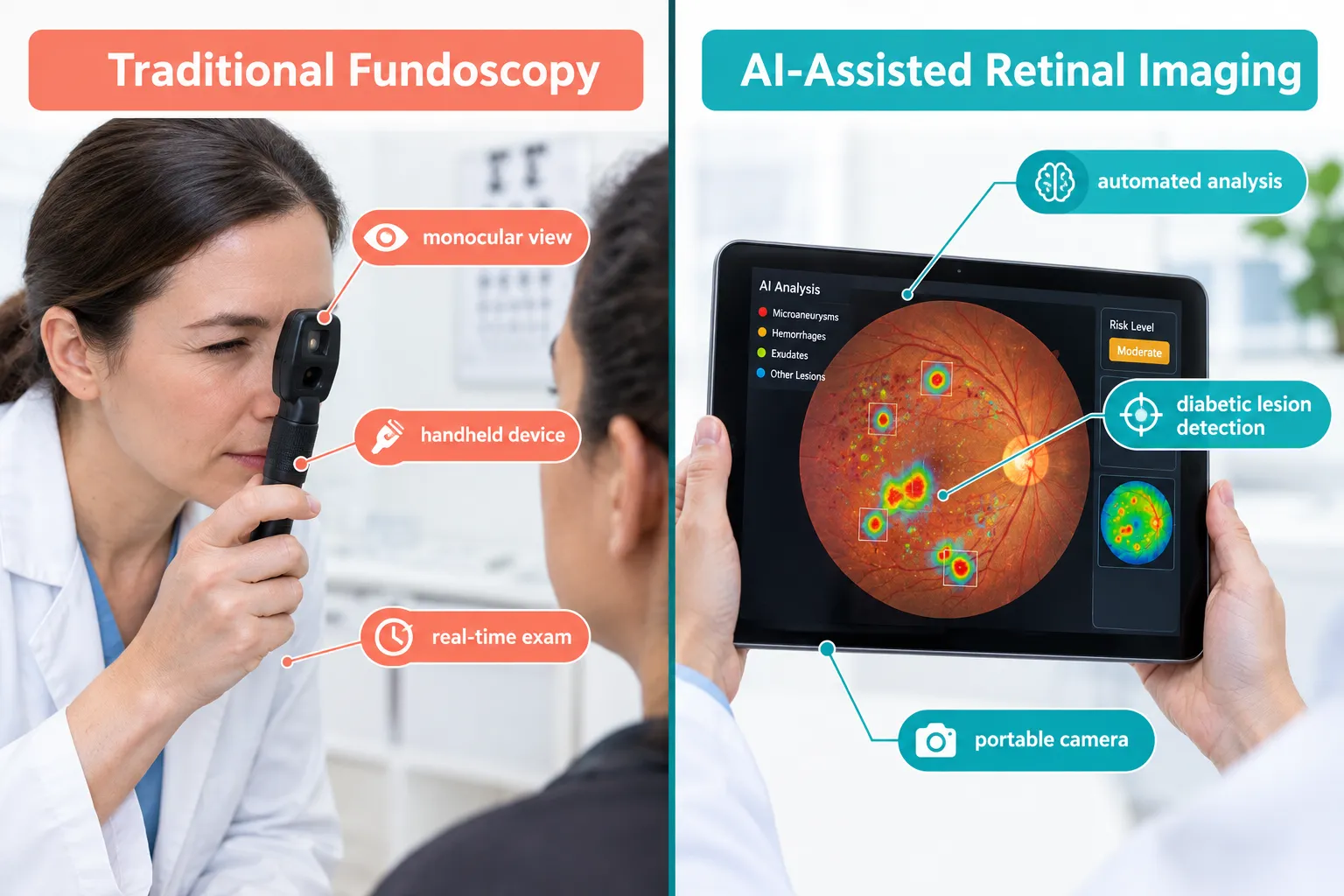
At the ENDO 2025 conference, researchers presented data on a mobile AI-powered retina tracker designed specifically for diabetic eye disease. The device uses a smartphone-compatible fundus camera paired with a deep-learning algorithm to screen for diabetic retinopathy in community and primary care settings, environments where traditional ophthalmoscopy resources are often limited.
For inherited retinal diseases (IRDs), a 2025 systematic review and meta-analysis found that AI models achieved high sensitivity and specificity when analyzing fundus images, outperforming traditional clinical grading in several categories. This is particularly significant because IRDs are rare and often go undiagnosed for years.
Perhaps most striking is the emerging use of retinal imaging for cardiovascular disease risk prediction. Research published in Frontiers in Cardiovascular Medicine demonstrated that AI analysis of fundus photographs can identify biomarkers predictive of major cardiac events, effectively turning a routine eye exam into a cardiovascular screening tool.
Main advantages of AI-enhanced fundoscopy:
- Consistent, reproducible grading that reduces human error
- Scalability for mass screening programs in underserved communities
- Real-time results in portable, low-cost formats
- Potential to detect systemic disease earlier than conventional methods
This broader paradigm, retinal imaging combined with AI as a clinical screening platform, represents one of the most exciting developments in preventive medicine in 2026. For patients interested in how personalized approaches to medicine are evolving, the article on personalized medicine vs. precision medicine provides valuable context.
Who Should Get Fundoscopy and How Often?
Not everyone needs annual fundoscopy, but certain patient populations should prioritize it as a routine part of their care.
High-priority groups include:
- People with type 1 or type 2 diabetes (annual exam recommended from diagnosis or within 5 years of type 1 onset)
- Adults over 50 with risk factors for AMD
- Patients with a family history of glaucoma
- Anyone with poorly controlled hypertension
- Patients on medications with known retinal toxicity (e.g., hydroxychloroquine)
Recommended screening frequency:
| Patient Profile | Recommended Frequency |
|---|---|
| Diabetes, no retinopathy | Annually |
| Diabetes, mild retinopathy | Every 6 months |
| Hypertension, uncontrolled | Annually or per specialist |
| Glaucoma risk | Annually |
| General adult (low risk) | Every 1-2 years after age 40 |
If you are unsure whether you need a referral to see a specialist, the guide on whether you need a referral to be seen by an endocrinologist can help clarify your next steps.
FAQs
Does fundoscopy hurt?
Fundoscopy is not painful. The examination itself involves a bright light that may feel temporarily uncomfortable, and if dilating drops are used, patients may experience mild stinging for a few seconds after application. The procedure is entirely non-invasive and typically takes 5 to 15 minutes once dilation is complete.
Can fundoscopy detect diabetes before I am diagnosed?
Fundoscopy can detect retinal changes caused by elevated blood sugar, but it is not used as a primary diabetes diagnostic test. Nonetheless, finding unexplained retinal microaneurysms or hemorrhages during a routine eye exam often prompts clinicians to order blood glucose testing, which can lead to an early diabetes diagnosis.
Is fundoscopy the same as a regular eye exam?
No. A standard vision or refraction exam checks your glasses prescription and basic eye health. A fundoscopy specifically examines the posterior segment of the eye, the retina, optic disc, and blood vessels, and is a more specialized component of a comprehensive eye examination. Many routine eye exams do not include a dilated fundus exam unless specifically requested or indicated.
How should I prepare for a fundoscopy appointment?
Bring sunglasses to wear after the exam, as your eyes will be light-sensitive for several hours if dilating drops are used. Arrange a ride if possible, since close-up vision (such as reading or driving) may be temporarily blurred. Inform your doctor of any eye medications you are currently using, and mention any personal or family history of eye disease, diabetes, or hypertension.
References
- American Academy of Ophthalmology. (2023). Diabetic Retinopathy Preferred Practice Pattern. https://www.aao.org/preferred-practice-pattern/diabetic-retinopathy-ppp
- National Eye Institute. (2022). Diabetic Retinopathy. U.S. Department of Health and Human Services. https://www.nei.nih.gov/learn-about-eye-health/eye-conditions-and-diseases/diabetic-retinopathy
- Centers for Disease Control and Prevention. (2023). Vision Health Initiative: Common Eye Disorders and Diseases. https://www.cdc.gov/visionhealth/basics/ced/index.html
- World Health Organization. (2023). Blindness and Vision Impairment. https://www.who.int/news-room/fact-sheets/detail/blindness-and-visual-impairment
- Teo, Z. L., et al. (2021). Global prevalence of diabetic retinopathy and projection of burden through 2045. Ophthalmology, 128(11), 1580-1591. https://doi.org/10.1016/j.ophtha.2021.04.027
Medical and Editorial Commitment
The content of this article is strictly for educational and informational purposes. It does not replace in-person professional medical diagnosis, advice, or treatment..
Read Privacy PolicyRelated Posts
11 Essential Strategies for Effective Diabetes Management
More than 38 million Americans are currently living with diabetes, yet studies consistently show that fewer than half meet their recommended blood [...]
What Are the Signs of Glaucoma? A Complete Guide to Symptoms, Risks, and When to Act
Over 3 million Americans are living with glaucoma right now, and roughly half of them do not know it. That is the [...]