
What if the first warning sign of osteoporosis appears long before pain or fractures ever do? In Bone Densitometry and Osteoporosis Detection Overview, we uncover how subtle changes inside your bones can reveal critical health insights. Guided by a trusted Bone Disease specialist in Queens, NY, this article explores the technology, timing, and unanswered questions that could change how you protect your bones—before it’s too late.
TL;DR
Bone densitometry (DXA) is a key, noninvasive tool for detecting early bone loss, diagnosing osteopenia and osteoporosis, and assessing fracture risk. The article explains patient preparation, positioning, DXA technology, and result interpretation using T-scores and Z-scores, highlighting how accurate measurement and regular follow-up help guide prevention, monitoring, and management of bone health over time.
Bone Disease specialist in Queens, NY
What Is Bone Densitometry Used for in Medical Diagnosis?
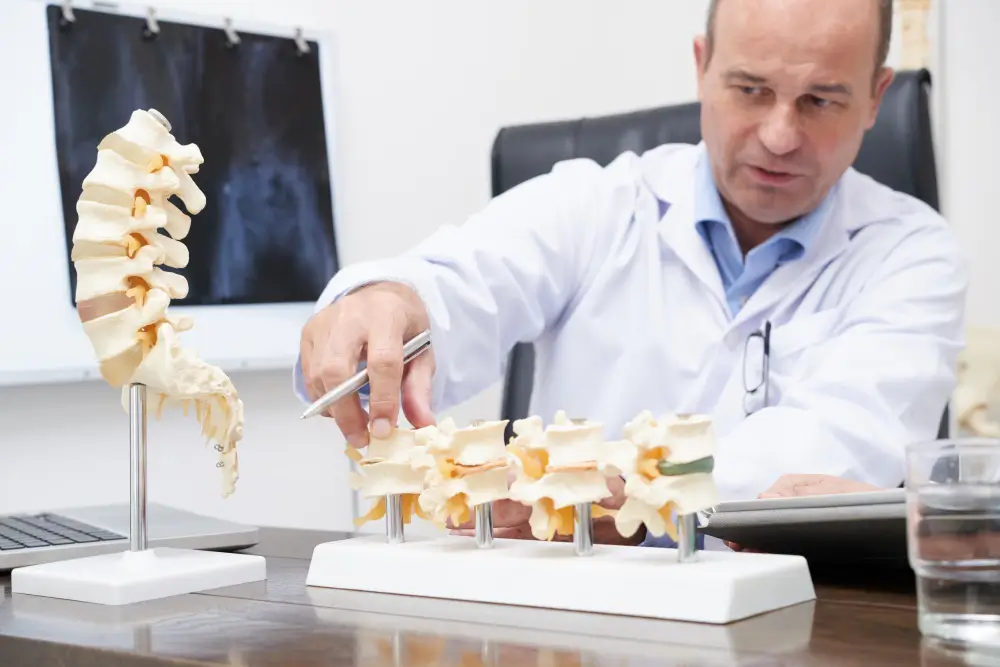
Bone densitometry, also known as DXA, is primarily used to assess bone health and support the clinical diagnosis of diseases like osteopenia and osteoporosis. This test allows the detection of bone mass loss in the early stages, even before symptoms or fractures appear.
From a medical perspective, densitometry is a highly useful tool for estimating fracture risk in patients with risk factors, such as postmenopausal women, older adults, those with a family history of osteoporosis, or individuals undergoing treatments that affect bone metabolism.
The exam is also crucial for monitoring patients undergoing osteoporosis treatment, as it helps evaluate the therapeutic response and track changes in bone mineral density over time, facilitating preventive clinical decisions.
Patient Preparation Before Bone Densitometry
Before performing a bone densitometry test, some basic guidelines should be followed to ensure accurate results and avoid interference during the exam. Preparation is straightforward, non-invasive, and designed to make the procedure quick, safe, and comfortable for the patient.
| Aspect | Instructions |
| Medications and supplements | Discontinue calcium, iron, multivitamins, and osteoporosis medications 24 to 48 hours before the exam. |
| Diet | No fasting is required. |
| Clothing | Wear comfortable clothes without metallic items such as zippers, buttons, buckles, or belts; a gown may be used in many cases. |
| Metal objects | Remove jewelry, watches, piercings, and non-removable dental appliances before the exam. |
| Previous studies | Inform if contrast studies, nuclear medicine, or CT scans were performed in the past two weeks. |
| Pregnancy | Notify if pregnant or possibly pregnant. |
Proper preparation contributes to smooth execution and reliable measurements. Following these simple guidelines and communicating relevant health history ensures a fast, painless, and non-invasive procedure lasting around 15 to 20 minutes.
Positioning and Equipment Setup for Accurate Scanning
The accuracy of bone densitometry largely depends on proper patient positioning and appropriate equipment setup. Correct alignment ensures clear images and reliable measurements of bone mineral density in the regions of interest, minimizing errors and avoiding interference during the study.
Patient Positioning and Equipment Configuration
The exam is performed on a wide table equipped with a mobile scanning arm and an X-ray generator located underneath. Before starting, patients are asked to remove any metallic items from the areas being examined, ensuring they are comfortable and still for optimal image acquisition.
Image Acquisition
- The operator confirms the positioning using the equipment’s optical system.
- The absence of artifacts that could distort the image is checked.
- Patient data, such as weight, height, and ethnicity, will be entered before proceeding with the scan.
- The osteodensitometer uses two energy sources to analyze bone mineral density.
Image Acquisition
The operator verifies positioning using the system’s optical alignment, checks for artifacts that may distort the image, and inputs patient data such as weight, height, and ethnicity before scanning begins.
The osteodensitometer uses dual-energy sources to analyze bone mineral density. Proper positioning and equipment configuration are vital for obtaining accurate bone density measurements and ensuring reliable results in osteoporosis or fracture risk assessment.
Measuring Bone Mineral Density With DXA Technology
Measuring bone mineral density using DXA (Dual-energy X-ray absorptiometry) technology quantifies bone mineral content in specific body regions, particularly the lumbar spine and hip. This technology provides consistent measurements that facilitate the technical evaluation of bone quality.
Objective and Procedure
- Objective: Quantify bone mineral content in specific anatomical regions for precise, reproducible bone mineral density measurements.
- Procedure: The patient lies on a table while a scanning arm moves over the region being studied, emitting two energy levels that distinguish bone tissue from soft tissue.
- Regions evaluated: Primarily the lumbar spine and hip, though other areas may be included based on technical indications.
- Duration: The study typically takes 10 to 30 minutes, depending on the number of regions analyzed.
- Exam Features: This quick, comfortable, non-invasive procedure allows serial measurements under consistent technical conditions.
DXA technology is known for providing highly reliable and consistent measurements, making it especially useful for comparing studies during follow-up and analyzing changes in bone mineral density over time.
Interpreting Scores to Identify Osteoporosis Risk
The interpretation of bone densitometry results primarily focuses on the T-score, which compares the patient’s bone mineral density with that of a young adult of the same sex. Based on this value, diagnostic criteria are established: a T-score of -1.0 or higher is considered normal, between -1.0 and -2.5 indicates osteopenia, and -2.5 or lower confirms osteoporosis. When osteoporosis is associated with one or more fragility fractures, it is classified as established osteoporosis.
A key factor in risk assessment is that the T-score reflects how many standard deviations the bone density is below the average young adult value. Each decrease of one standard deviation significantly increases fracture risk, multiplying it by 1.5 to 2 times. The most common measurement sites—lumbar spine, proximal femur, and radius—are used to establish the diagnosis, with the lowest value serving as the diagnostic criterion.
Additionally, the Z-score compares bone density with others of the same age, sex, and body size, particularly useful for younger populations to identify potential secondary causes of bone loss. A Z-score below -2.0 is considered below the expected range for age. Follow-up frequency is based on initial results: normal bone density is typically checked every 3 to 5 years, while osteopenia or osteoporosis requires more frequent evaluations, generally every 1 to 3 years, to assess progression and fracture risk.
Key Takeaways
- Bone densitometry (DXA) allows early detection of bone loss: DXA is a noninvasive exam used to evaluate bone health and identify osteopenia or osteoporosis at early stages. It can reveal subtle reductions in bone density before symptoms or fractures appear, enabling timely assessment and preventive clinical decisions.
- DXA is essential for diagnosis and fracture risk evaluation: The test supports clinical diagnosis by estimating fracture risk, particularly in individuals with known risk factors. It is also used to monitor changes in bone density over time, helping guide ongoing management and follow-up strategies.
- Proper patient preparation ensures accurate results: Simple preparation steps, such as avoiding calcium supplements and metal objects, help prevent interference with measurements. The exam does not require fasting and is designed to be quick, painless, and noninvasive for the patient.
- Correct positioning and equipment setup improve measurement reliability: Accurate patient alignment and proper use of scanning equipment are critical for obtaining clear images. Specific positioning techniques are applied depending on the body region being examined to ensure consistent and reliable bone density measurements.
- T-scores and Z-scores guide interpretation and follow-up planning: T-scores classify bone density status and estimate fracture risk, while Z-scores compare bone density with age-matched populations. Follow-up frequency is determined by these results, with closer monitoring required for osteopenia or osteoporosis.
FAQs
What type of doctor treats bone disease?
Bone diseases are treated by medical specialists who focus on bone health and metabolism. Depending on the condition, care may involve doctors trained in musculoskeletal, hormonal, or systemic disorders affecting bone strength and structure.
What kind of doctor is best for bone disorders?
The best doctor for bone disorders depends on the underlying cause of the condition. Specialists trained in bone metabolism, autoimmune conditions, or musculoskeletal health may be involved to diagnose, monitor, and manage bone density loss or structural changes.
What are the three major bone diseases?
Three common bone diseases include osteoporosis, osteopenia, and Paget’s disease of bone. These conditions can weaken bones, alter their structure, and increase the risk of fractures or deformities if not properly monitored.
Is it better to see a rheumatologist or endocrinologist for osteoporosis?
Both specialists treat osteoporosis, but the choice depends on the cause. Endocrinologists are often preferred for hormone-related or metabolic factors, while rheumatologists commonly manage cases linked to autoimmune disease or long-term steroid use.
Sources
- Ghasemi, N., Rokhshad, R., Zare, Q., Shobeiri, P., & Schwendicke, F. (2025). Artificial intelligence for osteoporosis detection on panoramic radiography: A systematic review and meta analysis. Journal of dentistry, 156, 105650.
https://www.sciencedirect.com/science/article/abs/pii/S0300571225000958
- Zhang, Y., Ma, M., Huang, X., Liu, J., Tian, C., Duan, Z., … & Geng, B. (2025). Machine learning is changing osteoporosis detection: an integrative review. Osteoporosis International, 1-14.
https://link.springer.com/article/10.1007/s00198-025-07541-x