
 By Atlantic Endo Medical Review Board
Pregnancy often raises important questions about medical tests and their safety. When thyroid changes or nodules appear during this stage, imaging becomes a common part of clinical evaluation. A thyroid ultrasound is widely recognized as a safe diagnostic tool during pregnancy because it uses sound waves rather than radiation. According to clinical evidence and professional guidelines, this exam helps physicians monitor thyroid health while protecting both mother and baby, especially when guided by an experienced Thyroid Specialist in Queens.
TL;DR
A thyroid ultrasound is considered a safe and effective imaging test during pregnancy. It relies on sound waves rather than radiation, which makes it suitable for evaluating thyroid nodules or changes without posing risks to the mother or developing baby. Medical guidelines support its use when clinically necessary to monitor thyroid health throughout pregnancy.
Care Options With a Thyroid Specialist in Queens
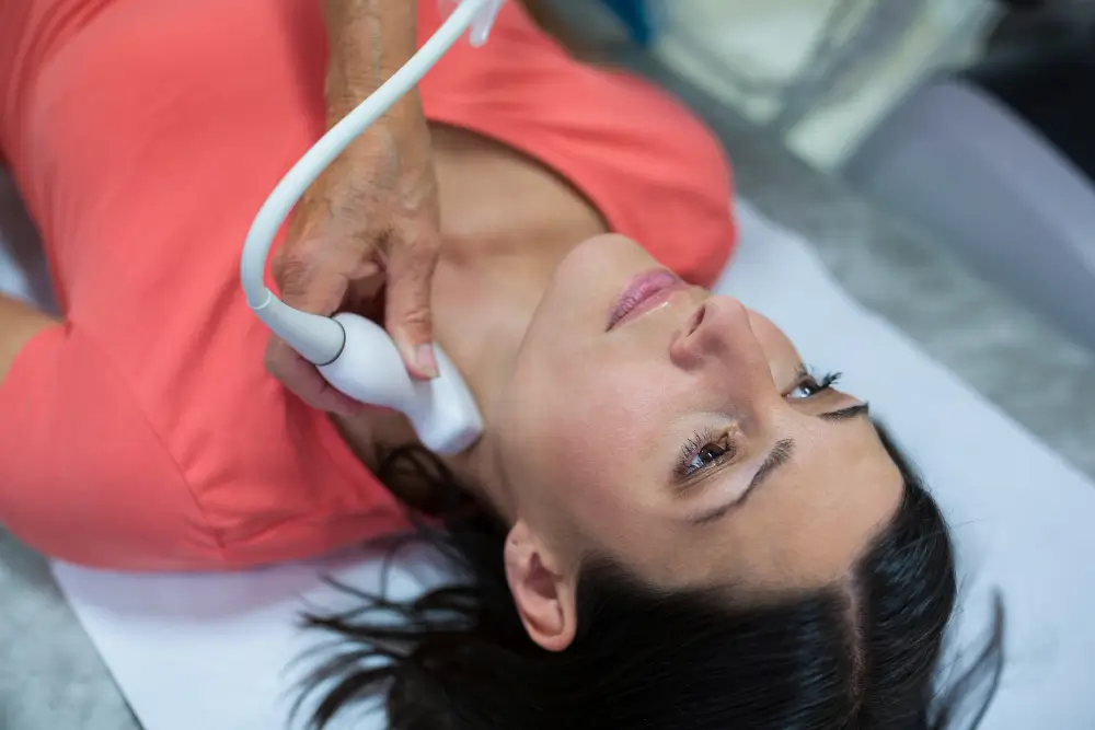
Table of Contents
How Does a Thyroid Ultrasound Work During Pregnancy?
Rather than relying on radiation, a thyroid ultrasound during pregnancy captures images of the thyroid gland using high-frequency sound waves. A small handheld probe sends these waves through the neck area, where they reflect off thyroid tissue and return as detailed images on a monitor. This technique allows healthcare professionals to observe nodules, cysts, or structural changes while maintaining a strong safety profile for pregnancy.
The procedure takes place with the patient lying comfortably and the neck slightly tilted back to improve visibility. A clear gel is applied to ensure smooth contact between the probe and the skin, helping produce accurate results. Clinical studies highlight that ultrasound offers real-time thyroid imaging, making it especially useful when physical exams or blood tests raise concerns during pregnancy.
Professional guidelines describe thyroid ultrasound as a key tool for assessing thyroid nodules safely in pregnant patients. The images support clinical judgment when deciding on follow-up, observation, or further evaluation. The exam remains painless, brief, and well tolerated, aligning with accepted standards for maternal and fetal care.
Purpose of Thyroid Ultrasound During Pregnancy
The main purpose of a neck gland ultrasound during pregnancy is to evaluate structural changes in the endocrine gland when clinical findings raise concern. This imaging exam allows healthcare professionals to assess nodules or gland enlargement using a radiation-free diagnostic method, which aligns with established safety standards for pregnant patients. Its role focuses on providing clarity when physical exams or blood tests suggest abnormalities that require closer evaluation.
- Detect thyroid nodules and measure their size
- Assess the shape and internal features of thyroid tissue
- Distinguish solid findings from fluid-filled structures
- Support monitoring when changes are observed over time
- Help identify characteristics linked to higher clinical risk
- Guide decisions about follow-up or additional testing
- Assist in determining whether biopsy should be considered
Overall, this imaging approach plays an important role in clinical assessment during pregnancy by offering detailed anatomical information without added risk. The findings help clinicians make informed decisions while maintaining a cautious and evidence-based approach to maternal care. Its reliability and safety explain why it remains a preferred option when thyroid evaluation is needed during pregnancy.
How the Ultrasound Procedure Is Performed
During pregnancy, the exam is carried out as a straightforward, noninvasive imaging procedure focused on examining the neck area. The patient lies comfortably on an exam table with the neck gently extended to improve visibility. Medical guidelines describe the process as safe, painless, and well tolerated, with no recovery time required. The objective is to obtain clear anatomical images while prioritizing comfort and overall safety.
| Step | What Happens |
| Patient positioning | The patient lies comfortably with the neck gently tilted back |
| Gel application | A clear gel is applied to improve sound wave transmission |
| Probe movement | A handheld device is moved across the neck area |
| Image capture | Real-time images of thyroid structure appear on a monitor |
| Duration | The exam typically lasts 15 to 30 minutes |
After the exam, no recovery time is needed and normal activities can be resumed immediately. The images obtained help clinicians evaluate thyroid structure and guide medical decisions when necessary. Professional recommendations emphasize this procedure as a reliable option within pregnancy-safe diagnostic imaging, supporting careful assessment without added risk.
Safety Considerations for the Mother and Baby
Safety is a primary concern when any diagnostic test is performed during pregnancy. Medical evidence shows that sound wave–based imaging does not involve ionizing radiation, making it appropriate for evaluating gland-related conditions without exposing the mother or developing baby to unnecessary risk. Professional guidelines consistently recognize this method as compatible with pregnancy when imaging is clinically indicated.
From a maternal perspective, the procedure is considered noninvasive and well tolerated, requiring no medications, injections, or recovery time. It does not interfere with normal pregnancy care and can be performed at any trimester when needed. Research highlights its usefulness in evaluating gland structure while maintaining a strong safety profile throughout pregnancy.
For fetal safety, clinical studies and expert recommendations emphasize that sound wave–based imaging has no known harmful effects on fetal development. The absence of radiation exposure supports its continued use when thyroid evaluation is necessary. This approach allows healthcare professionals to balance accurate diagnosis with careful protection of both mother and baby.
Situations When a Thyroid Ultrasound Is Recommended
This imaging exam is recommended during pregnancy when clinical findings suggest possible thyroid changes that require closer evaluation. Medical literature and professional guidelines emphasize its use when physical examination or laboratory results indicate abnormalities. The goal is to obtain clear anatomical information while following established safety standards for pregnant patients and supporting accurate clinical assessment.
- Presence of a palpable neck mass or glandular enlargement
- Detection of gland nodules during physical examination
- Abnormal endocrine function test results that raise structural concerns
- History of gland-related disease requiring closer monitoring
- Rapid growth or change in a previously identified nodule
- Clinical features associated with higher suspicion of malignancy
- Need to guide decisions related to observation or further evaluation
In these situations, this imaging method supports evidence-based clinical decision-making by providing detailed structural insight. The findings help determine whether continued monitoring or additional diagnostic steps are appropriate. Its safety profile and diagnostic value explain why it remains a preferred option when thyroid evaluation is necessary during pregnancy.
Interpreting Results and Next Medical Steps
Once the imaging findings are available, attention is directed toward understanding what the observed features mean in a clinical context. Size, structure, and internal composition are reviewed to determine whether findings appear typical or suggest the need for closer evaluation. Research shows that most findings identified during pregnancy are benign and can be assessed using standardized imaging criteria.
When nodules or structural changes are detected, clinicians consider factors such as growth behavior and visual characteristics. Findings associated with lower clinical concern often lead to a monitoring approach rather than immediate intervention. This strategy allows careful observation while minimizing unnecessary procedures during pregnancy.
If certain features raise concern, the next medical steps are selected cautiously. Professional guidelines recommend individualized management that balances diagnostic clarity with maternal and fetal safety. This may include referral, follow-up imaging, or further evaluation when appropriate, following a conservative, evidence-based approach.
Key Takeaways
- Ultrasound-based imaging is considered safe during pregnancy
It uses sound waves instead of radiation, which makes it appropriate for evaluating thyroid changes while protecting both the mother and the developing baby. - The procedure is simple, noninvasive, and well tolerated
It requires no medications, injections, or recovery time and can be performed at any stage of pregnancy when clinically indicated. - Imaging helps assess thyroid structure and guide care decisions
It allows clinicians to evaluate nodules, gland size, and internal features to determine whether monitoring or further evaluation is needed. - Most findings during pregnancy are benign and managed conservatively
Standardized imaging criteria help identify low-risk features, often leading to observation rather than immediate intervention. - Results support individualized, evidence-based medical planning
Findings guide follow-up, referrals, or additional testing when necessary, always prioritizing maternal and fetal safety.
FAQs
Why Would You Need To See A Thyroid Specialist?
You may need a thyroid specialist when exams or imaging show nodules, enlargement, or changes that require focused evaluation. These professionals help interpret findings and guide safe, appropriate next steps.
Who Is the Best Doctor To See For Thyroid Problems?
An endocrinologist is usually the best option for thyroid conditions, as they specialize in hormone-related disorders. Care may also involve other physicians depending on the clinical findings.
What Lifestyle Causes Thyroid Problems?
Chronic stress, poor sleep, unbalanced nutrition, and excessive iodine intake can affect thyroid function. Lifestyle factors may worsen symptoms rather than directly cause disease.
What Are 5 Foods To Avoid For Thyroid?
Highly processed foods, excess soy, large amounts of raw cruciferous vegetables, iodine-rich foods, and refined sugars may need moderation depending on thyroid health.
What Habits Hurt Your Thyroid?
Smoking, chronic stress, irregular sleep, skipping meals, and unsupervised supplement use may negatively impact thyroid health over time.
Sources
- Marzouk, M., Papaleontiou, M., et al. (2019).
Thyroid nodules and cancer during pregnancy, postpartum and imaging modalities.
PubMed Central (PMC).
https://pmc.ncbi.nlm.nih.gov/articles/PMC7242146/ - American Thyroid Association. (2011).
Guidelines of the American Thyroid Association for the diagnosis and management of thyroid disease during pregnancy and postpartum.
American Thyroid Association.
https://pmc.ncbi.nlm.nih.gov/articles/PMC3472679/
You May Also Like:
- Reasons to See an Endocrinologist as Soon as Possible
- What to Expect at Your First Endocrinologist Appointment for Thyroid
- Should I See an ENT or Endocrinologist for Thyroid?
Medical and Editorial Commitment
The content of this article is strictly for educational and informational purposes. It does not replace in-person professional medical diagnosis, advice, or treatment..
Read Privacy PolicyRelated Posts

How to Enhance Your Metabolism: Step by Step in Rego Park, NY
Arrange a Visit Semaglutide Injections and IV Therapy in Rego Park, NY. Nearly 40% of American adults are living with obesity, yet [...]
What Organ Is Most Affected by Glucose?
While every cell in your body relies on sugar, one vital organ bears the brunt of unregulated glucose levels, often suffering in [...]