
 By Atlantic Endo Medical Review Board
A routine breathing test might reveal more than you think—but can it actually help detect lung cancer early? Many patients visiting a pulmonary doctor in Queens assume pulmonary function tests are purely about measuring lung capacity. Yet some specialists suggest these tests may hint at deeper issues. So what can they truly uncover—and what might they miss? The answer may surprise you.
TL;DR
Pulmonary function tests measure how well the lungs work by evaluating airflow, lung capacity, and gas exchange, but they cannot directly detect lung cancer. Their main role is to identify breathing disorders, monitor respiratory health over time, and assess treatment response. For early lung cancer detection, the recommended method is low-dose CT scanning, which can identify small lung nodules in high-risk individuals.
Consult a pulmonary doctor in Queens
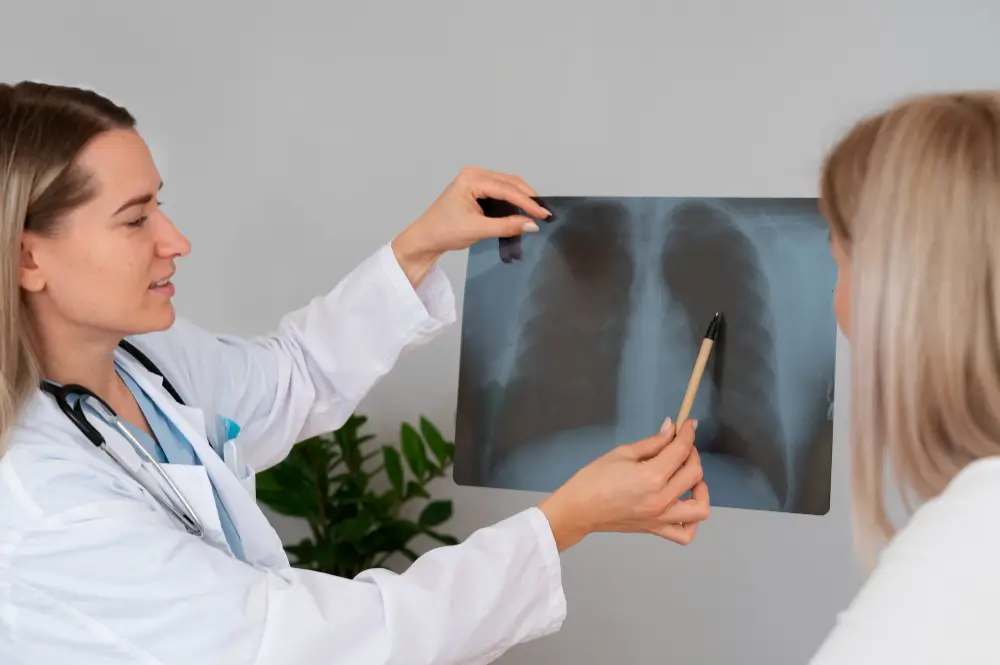
Table of Contents
How Accurate Are Pulmonary Function Tests for Early Lung Cancer?
Pulmonary function tests are not the primary method used to detect lung cancer in its early stages. These tests evaluate functional changes in breathing, such as lung capacity and airflow, but they do not identify tumors or specific nodules. Because of this, their ability to diagnose early lung cancer is limited.
Tests such as spirometry can detect respiratory problems including COPD or restrictive lung patterns. These conditions may represent risk factors linked to lung disease, but they do not confirm the presence of a tumor. Their main role is evaluating overall lung function.
Low-dose computed tomography (LDCT) is considered the standard method for early lung cancer detection because it can identify small pulmonary nodules. This test is recommended for individuals at high risk, such as adults ages 50–80 with a history of smoking. Detecting cancer in stage 1 can raise five-year survival rates to nearly 90 percent.
Assessing Lung Capacity and Airflow
Evaluating lung capacity and airflow helps explain how the lungs move air in and out of the body. These tests measure how much air the lungs can hold and how quickly it moves during breathing. Several non-invasive exams help specialists identify ventilation abnormalities and evaluate how the respiratory system functions mechanically.
| Test | What It Evaluates | Clinical Purpose |
| Spirometry | Measures the volume of air inhaled and exhaled and airflow speed | Detects obstructive or restrictive breathing patterns |
| Body Plethysmography | Calculates total lung capacity and residual lung volume | Determines how much air remains in the lungs after exhalation |
| Diffusing Capacity (DLCO) | Measures how well oxygen passes into the bloodstream | Evaluates gas exchange efficiency |
| Exercise Testing | Measures respiratory performance during physical activity | Evaluates lung response to exertion |
These tests provide a broad view of lung performance by analyzing ventilation, lung volumes, and oxygen transfer. With this information, healthcare professionals can detect respiratory abnormalities and observe how the lungs manage airflow during breathing.
Measuring Breathing Efficiency Over Time
Pulmonary function tests can also track breathing performance over time. By repeating certain measurements periodically, specialists can determine whether respiratory function remains stable, improves, or shows gradual change.
Measurements Used to Track Breathing Efficiency
- FEV1 (Forced Expiratory Volume in 1 second): Amount of air expelled during the first second of forced exhalation.
- FVC (Forced Vital Capacity): Total volume of air exhaled during a deep, complete breath.
- Minute Ventilation: Total air moving in and out of the lungs in one minute.
- Maximum Voluntary Ventilation (MVV): Measures ventilatory capacity while breathing rapidly and deeply for a short period.
- Forced Expiratory Flow (FEF 25–75%): Average airflow during the middle portion of exhalation.
Parameters Used to Interpret the Results
| Parameter | What It Measures | General Interpretation |
| FEV1/FVC Ratio | Relationship between airflow speed and total exhaled volume | Values above about 0.70 are generally considered normal in adults |
| Predicted Values | Comparison with standardized reference values | Based on age, height, sex, and population data |
| Ventilation Measurements | Overall breathing capacity | Identifies variations in respiratory efficiency |
Tracking these parameters over time helps specialists identify trends in respiratory performance and evaluate how the lungs respond to activity or other conditions.
Identifying Abnormal Respiratory Patterns
Pulmonary function tests, especially spirometry, help identify abnormal breathing patterns by analyzing airflow during inhalation and exhalation. Interpreting these patterns helps detect respiratory conditions and guide clinical evaluation.
| Respiratory Pattern | Characteristic | Associated Conditions |
| Obstructive | Reduced airflow due to narrowed airways | Asthma, chronic bronchitis, emphysema |
| Restrictive | Reduced lung expansion and lower lung volumes | Pulmonary fibrosis, sarcoidosis, severe obesity |
| Functional | Irregular breathing mechanics | Hyperventilation, thoracic breathing |
Findings in the Flow-Volume Curve
Flow-volume curves may also reveal irregularities during testing:
- Negative flows: May indicate air leakage during the test.
- Cough artifacts: Produce irregular spikes in the curve.
- Irregular descending curve: May reflect poor technique or inconsistent patient effort.
Recognizing these patterns allows clinicians to interpret pulmonary test results more accurately and identify possible breathing abnormalities.
Comparing Results with Previous Tests
Comparing current pulmonary function test results with previous evaluations helps detect changes in respiratory function over time. This analysis shows whether breathing remains stable, improves, or declines.
Reviewing tests performed at different times can reveal changes that may not appear in a single evaluation.
- Disease progression: Tracks the development of chronic respiratory diseases.
- Treatment effectiveness: Evaluates whether therapy improves lung function.
- Rapid decline detection: Identifies significant reductions in respiratory performance.
- Condition monitoring: Shows whether lung function remains stable or changes.
When reviewing results, specialists often focus on key indicators:
- FEV1: Measures air expelled during the first second; decreases may indicate worsening obstruction.
- FVC: Total air exhaled after deep inhalation; lower values may suggest reduced lung capacity.
- Lung volumes: Help identify changes in obstructive or restrictive patterns.
Reliable comparisons require consistent testing technique and similar conditions so that differences reflect real changes in lung function.
Supporting Diagnosis Alongside Imaging
Pulmonary function tests such as spirometry are often used with lung imaging studies, including chest X-rays and CT scans, to evaluate both lung function and lung structure.
Pulmonary tests measure breathing capacity, airway obstruction, and gas exchange, while imaging studies provide visual information about lung anatomy. Chest X-rays can reveal inflammation, fluid, tumors, or calcifications, while CT scans provide detailed images that help identify conditions such as fibrosis or emphysema in earlier stages.
Combining these evaluations helps confirm respiratory diseases, measure their severity, and monitor treatment response. This approach also supports disease monitoring, pre-surgical evaluation, and identification of lung damage related to environmental or occupational exposure.
Key Takeaways
- Pulmonary function tests do not directly detect lung cancer: These tests evaluate airflow, lung capacity, and gas exchange but cannot identify tumors or lung nodules.
- Low-dose CT scans are the recommended method for early detection: This imaging technique can detect small lung nodules and is commonly recommended for high-risk individuals.
- Pulmonary function tests measure how well the lungs work: Exams like spirometry and diffusion tests analyze airflow, lung volumes, and oxygen transfer during breathing.
- Repeated testing helps track respiratory changes over time: Comparing results such as FEV1 and FVC allows specialists to monitor disease progression and treatment response.
- Combining functional tests with imaging improves diagnosis: Pulmonary tests assess lung function, while imaging studies reveal structural changes in lung tissue.
Sources
- Ponce, M. C., Sankari, A., & Sharma, S. (2023). Pulmonary function tests. In StatPearls [internet]. StatPearls Publishing.
https://www.ncbi.nlm.nih.gov/sites/books/NBK482339
- Kakavas, S., Kotsiou, O. S., Perlikos, F., Mermiri, M., Mavrovounis, G., Gourgoulianis, K., & Pantazopoulos, I. (2021). Pulmonary function testing in COPD: looking beyond the curtain of FEV1. NPJ primary care respiratory medicine, 31(1), 23.
https://www.nature.com/articles/s41533-021-00236-w
FAQs
What does a pulmonary doctor do?
A pulmonary doctor, also known as a pulmonologist, specializes in diagnosing and treating diseases of the respiratory system, including the lungs, airways, and breathing muscles. They manage conditions such as asthma, COPD, infections, sleep apnea, and lung cancer. To evaluate lung health, they may use specialized tests and develop treatment plans that help patients breathe more comfortably.
What four questions will you ask to a pulmonologist?
When visiting a pulmonologist, it is helpful to ask questions that clarify your condition and treatment plan. Common questions include: what is my specific diagnosis and its cause, what treatment options are available and their goals, what lifestyle changes I should make, and what warning signs I should watch for. These questions help patients better manage their respiratory health.
What’s the difference between a pulmonary doctor and a lung doctor?
There is no difference between a pulmonary doctor and a lung doctor. Both terms refer to the same type of medical specialist, commonly called a pulmonologist, who focuses on diagnosing and treating respiratory conditions. These specialists receive advanced training to manage diseases affecting the lungs and airways, including asthma, COPD, sleep apnea, and lung cancer.
Can a pulmonary doctor help with asthma?
Yes, a pulmonary doctor can help diagnose and manage asthma by evaluating lung function and identifying triggers that affect breathing. They may recommend treatments, medications, and lifestyle adjustments to control symptoms and improve respiratory health. Their goal is to help patients maintain stable breathing and prevent asthma flare-ups over time.
Medical and Editorial Commitment
The content of this article is strictly for educational and informational purposes. It does not replace in-person professional medical diagnosis, advice, or treatment..
Read Privacy PolicyRelated Posts

Treatment for Neuropathy in Legs and Feet: A Complete Guide for Patients Seeking in Queens, NY
Last updated: June 11, 2026 Quick Answer: Diabetic neuropathy in the legs and feet is nerve damage caused by chronically high blood [...]

Vitamin D3 vs D2 for Bone Health: Which Is Better? A Guide to Bone Specialist Services
Last updated: June 4, 2026 Quick Answer: Vitamin D3 (cholecalciferol) is the superior choice for bone health. It raises and maintains blood [...]